



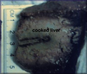
Gross in vitro liver specimen showing heated, dead liver in the middle with uncooked normal liver on the outside. Note the sharp predictable margin between treated and untreated. The dark line in the middle is the needle tract.
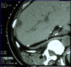
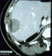
This 3cm hepatocelluar carcinoma appeared elsewhere in the liver after prior surgery for the same disease. Repeat surgery was avoided by treating this tumor by inserting a needle through the skin and killing all of the tumor. Note that there is a clean margin of dark and devascularized, dead tissue including the old tumor and the surrounding tissue.
Colon carcinoma liver metastasis ▾
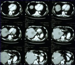
CT scan of the liver before ablation shows large solitary lesion from colorectal carcinoma.
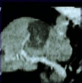
Coronal reconstructed image after treatment shows dark treated area without contrast enhancement in the area of the tumor. The dark area is coagulative necrosis, or dead cells.
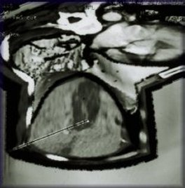
Three-dimensional reconstruction after ablation.
This colon cancer metastasis to the liver had been previously treated with surgery and cryotherapy but had recurred and failed chemotherapy.
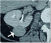
Pre-treatment scan of kidney tumor
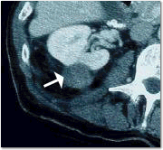
Post-treatment CGT scan of kidney tumor showing complete treatment, seen as eradication of contrast enhancement
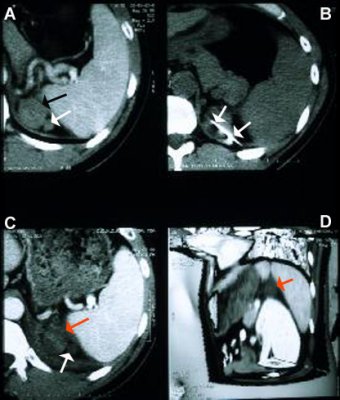
A: Pre-treatment enhancing adrenocortical carcinoma
B: RFA treatment needle in tumor during treatment
C: After RFA, there is no enhancement in tumor, consistent with complete tumor death
D: 3D with planes cut away showing treated tumor with close proximity of kidney, pancreas, spleen and stomach, without collateral damage