Content on this webpage is provided for historical information about the NIH Clinical Center. Content is not updated after the listed publication date and may include information about programs or activities that have since been discontinued.

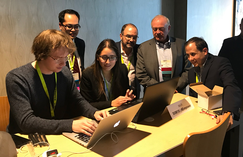
In June, two researchers from the NIH Clinical Center collaborated with external colleagues to win first place in a 'Hackathon' at the Society of Informatics Imaging in Medicine annual conference – a conference that helps accelerate Artificial Intelligence (AI) in medicine. The team, called PACS-Man (PACS stands for Picture Archiving and Communication System), included the NIH Clinical Centers' Dr. Les Folio, lead computed tomography radiologist in the Department of Radiology and Imaging Sciences, and Dr. Michael Do, Intramural Research Training Award (IRTA) and Folio's postdoctoral research fellow in the department.
The Hackathon is a collaborative effort between informaticists, technologists and clinicians to improve upon existing healthcare IT systems and develop innovative solutions to challenging issues in healthcare IT today. With a merging of developers and clinicians, team PACS-Man received first place for its concept and platform to complete the mundane task of tumor quantification, or measurement, and provide annotations, or measurements/drawings in images, to better train AI algorithms. Crowdsourcing the capability to perform quantification on imaging is emerging as an option to accommodate the need for labeled image datasets for "supervised" learning of AI algorithms.
Team PACS-Man embodied this in their winning project/proof of concept by successfully developing a platform that was easily accessible on any mobile device to annotate and segment normal anatomical structures on CT images. To further encourage user participation, they made it into a type of game by providing a leader scoreboard to depict the precision and accuracy of annotations/segmentations made by each participant.
- Molly Freimuth

